State of the Art Pediatrics: The Connectome: A New Imaging Dimension to Map Children’s Brains
Column Author: Avner Meoded, MD | Associate Professor of Radiology
Matthew Genchev, MD | Research Scholar
Column Editor: Amita R. Amonker, MD, FAAP | Assistant Professor of Pediatrics, University of Missouri-Kansas City School of Medicine; Clinical Assistant Professor of Pediatrics, University of Kansas School of Medicine
The field of clinical neuroradiology is largely concerned with the diagnosis of neurologic diseases using mainly conventional imaging modalities. While such techniques as CT scans, MRIs and X-rays are clinically valuable and robust in identifying the underlying causes of patient symptoms and diagnoses, they are not — alone — sufficient for the full characterization of network-based diseases. These connectopathies result from altered, lost or gained connections between functional centers within the brain — connections that traditional modalities are inherently unable to delineate.
As such, the advanced neuroimaging lab at Children’s Mercy Hospital, headed by Dr. Avner Meoded — along with the neuroradiology team — is using several new and emerging techniques and technologies to extend the reach of conventional imaging methods, generating active research, and improving outcome for children affected by neurologic disorders.
Central to this effort is the use of MRI, specifically, diffusion weighted/tensor imaging (DWI/DTI), a non-invasive neuroimaging technique capable of depicting the wiring diagram of children’s brain. In clinical practice, DTI derived images are directly interpreted by a neuroradiologist for their qualitative properties. However, DTI can be augmented by a process known as fiber tractography to create a three-dimensional reconstruction of nerve tracts within the brain.1 (Figure 1B) This reconstruction shows the neural pathways between brain centers, but it does not show which areas of the brain are connected. Therefore, fiber tractography is then correlated with a parcellated anatomical image of the individual patient’s brain (Figure 1A) to determine the overall structural connectivity of the patient.2,3
This process results in the creation of a patient connectome. A connectome is the “comprehensive structural description of the network of elements and connections forming the human brain.”2 How a connectome is represented is variable (Figure 1C), but basically it displays how functional or structural units of the brain (elements/nodes) are connected through axonal fiber tracts (connections/edges). This display provides a view of brain connectivity that is hidden in traditional imaging.
Leveraging the connectome opens a new approach for analyzing and treating diseases: connectomics. In the clinical research setting, connectomics has been successfully applied to better understand disease pathways, apply differential diagnoses, provide prognoses, and even dictate treatment type and efficacy.4 As in the case of congenital brain malformations like agenesis of the corpus callosum (AgCC), connectome analysis has proven useful in identifying alternative network hubs — highlighting neural plasticity in patients who are unable to send interhemispheric signals.3 While AgCC is possible to visualize with traditional methods, a connectomic view shows how the disease affects the brain as a whole organizational unit. This characterization draws from the connectome’s strength in providing quantitative measures of global and local connectedness using network statistics. Whereas traditional neuroradiology relies on qualitative observation, connectomics can provide a more concrete and numerical measure for physicians and researchers. This approach can go beyond congenital malformations and has been useful in clinical evaluation of autism spectrum disorder, attention-deficit/hyperactivity disorder, schizophrenia, epilepsy, neurooncology, ischemia and more.3,4
At the advanced neuroimaging lab, research targeted toward understanding pediatric epilepsy through a connectomic modality is underway. Epilepsy is commonly considered a network disease and presents one of the most interesting entity suitable for modern network analysis. As such, surgical target definition, treatment outcome prediction, and diagnosis of this inherently network-based disease are all likely to benefit from connectomic analyses that move research approaches from a focus-detection paradigm to a global network characterization.
However, the ultimate aim this approach is to take connectomics beyond the research sphere and into the clinical setting. Collaborations between the advanced research computing (ARC) team and the advanced neuroimaging lab are underway to develop a pipeline for the construction of connectomes at Children’s Mercy to increase the availability for both clinicians and researchers. While connectomics hasn’t made its official entry into the diagnostic and clinical sphere, this effort will put Children’s Mercy at the forefront of clinical research and the creation of a world of wellbeing for all children.
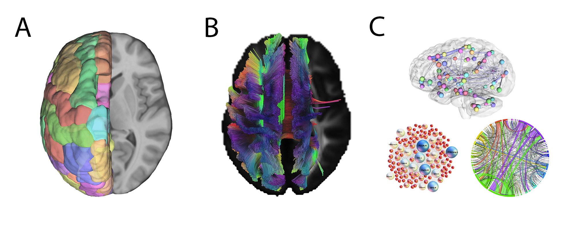
Figure legend
Figure 1: A) Axial view of a T1-weighted anatomical image of the brain with partially overlaid parcellated regions from a standard atlas. B) Axial view of a DTI image of the brain overlaid with partial fiber tracts derived from deterministic tractography. C) Various brain network representations including a sagittal anatomical view of a network, a network with highly connected nodes enhanced, and a classic connectogram.
References:
- Meoded A, Orman G, Huisman T. Diffusion weighted and diffusion tensor MRI in pediatric neuroimaging including connectomics: principles and applications. Semin Pediatr Neurol. 2020;33:100797.
- Meoded A, Huisman T, Casamassima MGS, Jallo GI, Poretti A. The structural connectome in children: basic concepts, how to build it, and synopsis of challenges for the developing pediatric brain. Neuroradiology. 2017;59:445-460.
- Meoded A, Goldenberg NA, Huisman T. Structural connectomics: state of the art and applications in pediatric neurodevelopmental disorders, neuro-oncology, and arterial ischemic stroke. J Pediatr. 2020;221S:S37-S42.
- 4. Damoiseaux JS, Altmann A, Richiardi J, Sadaghiani S. Chapter 21 - Applications of MRI connectomics. In: Choi I-Y, Jezzard P, eds. Advances in Magnetic Resonance Technology and Applications. Academic Press; 2021:323-338.