Visual Diagnosis: Thickened and Discolored Toenails
Column Author: Sean Reynolds, MBBCH | Associate Program Director, Pediatric Dermatology Fellowship, Assistant Professor of Pediatrics, University of Missouri-Kansas City School of Medicine; Clinical Assistant Professor of Internal Medicine, University of Kansas School of Medicine
Column Editor: Angela L. Myers, MD, MPH | Chief Wellbeing Officer, Center for Wellbeing, Professor of Pediatrics, University of Missouri-Kansas City School of Medicine; Clinical Assistant Professor of Pediatrics, University of Kansas School of Medicine
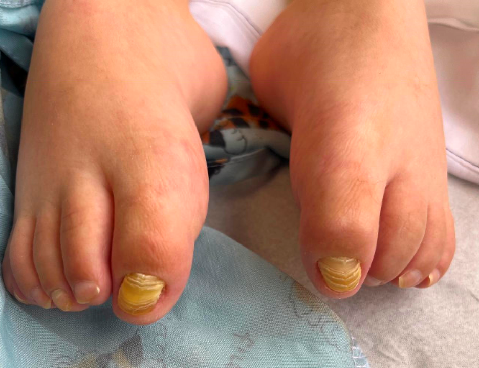
A 9-year-old boy with a history of cerebral palsy and seizures presents for evaluation of changes in his big toenails. His mother reports that his big toenails have looked different since early childhood but that the thickening and discoloration have worsened over the past couple of years. They deny any recent rash. He has had several ingrown toenails but has never developed paronychia. On examination, he has thickening and a yellow-brown discoloration of the bilateral great toenails. The remainder of his toenails and his fingernails appear normal. There is no periungual erythema or scale. A dermatophyte culture was performed on a clipping of the toenail and was negative.
Which of the following is the most likely diagnosis?
- Onychomycosis
- Congenital malalignment of the great toenails
- Nail psoriasis
- Onychomadesis
- Congenital malalignment of the great toenails
The patient presents with a history of nail changes isolated to the bilateral great toenails. The changes have been present since early childhood.
On physical examination, the bilateral great toenails show lateral deviation of the nail plates as well as yellowing and thickening (hyperkeratosis).
These features in the context of otherwise normal-appearing toenails and fingernails along with a history of recurrent ingrown toenails are consistent with a clinical diagnosis of congenital malalignment of the great toenails.
Congenital malalignment of the great toenails is a condition in which the nail matrix of the great toenail is laterally deviated and can lead to associated nail dystrophy of the affected toenails. It can be inherited in an autosomal dominant fashion.
Clinical Features:
- The lateral deviation of the nail matrix results in lateral deviation of the nail plate relative to the longitudinal axis of the distal phalanx. This can result in longitudinal ridging, thickening of the nail plate and discoloration of the nail plate.
- It is typically present at birth or early childhood but acquired forms manifesting later in life have been reported.
- While milder cases can self-resolve over time, the condition can be complicated by recurrent ingrown toenails, paronychia (inflammation of the proximal and lateral nailfolds) and onychogryphosis (“ram’s horn” nail deformity) of the affected nails.
The diagnosis is made clinically by recognizing the lateral deviation of the nail plates isolated to the bilateral great toenails. When dystrophic nail changes have developed (such as yellowing and hyperkeratosis), they are typically isolated to the great toenail plates with otherwise normal toenails and fingernails.
Onychomycosis: While secondary dermatophyte infection of congenital malalignment of the great toenails can occur, it would be unusual for symmetric primary onychomycosis of the bilateral great toenails to occur without involvement of other toenails or fingernails. In addition, a dermatophyte culture was performed in this case and was negative. The lateral deviation of the nail plate would not be expected to result from onychomycosis.
Psoriasis: While psoriasis can lead to symmetric nail dystrophy, it typically involves multiple fingernails and/or toenails, not just isolated to the great toenails. The typical findings of nail pitting, distal onycholysis and oil spotting are not seen in this case. In addition, nail psoriasis would not typically cause lateral deviation of the nail plate.
Onychomadesis: This condition refers to the proximal separation of the nail plate from the nail matrix, secondary to a temporary interruption in nail plate growth. Clinically, the proximal and distal nail plates are discontinuous. The temporary cessation in nail plate growth can be due to infections (most commonly weeks to months after hand, foot and mouth disease in children), inflammatory causes, and certain medications. It more commonly affects the fingernails than the toenails; involvement isolated to the great toenails would be unusual and would not be expected to cause lateral displacement of the nail plate.
Treatment:
If noted in infancy and the degree of deviation is severe, the deviation may be corrected surgically. However, surgery is typically only effective when patients are less than 2 years of age. Some cases will self-correct over time, and many parents may prefer observation. If complications develop in older children, such as recurrent ingrown toenails or recurrent paronychia, then typically nail avulsion with matriectomy (with resultant absence of the great toenails) is necessary for definitive treatment. Simple removal of the nail usually results in recurrence as it does not correct the deviated nail matrix.