What’s the Diagnosis?: Dermatology
Column Author and Editor: Sean Reynolds, MBBCH | Associate Program Director, Pediatric Dermatology Fellowship
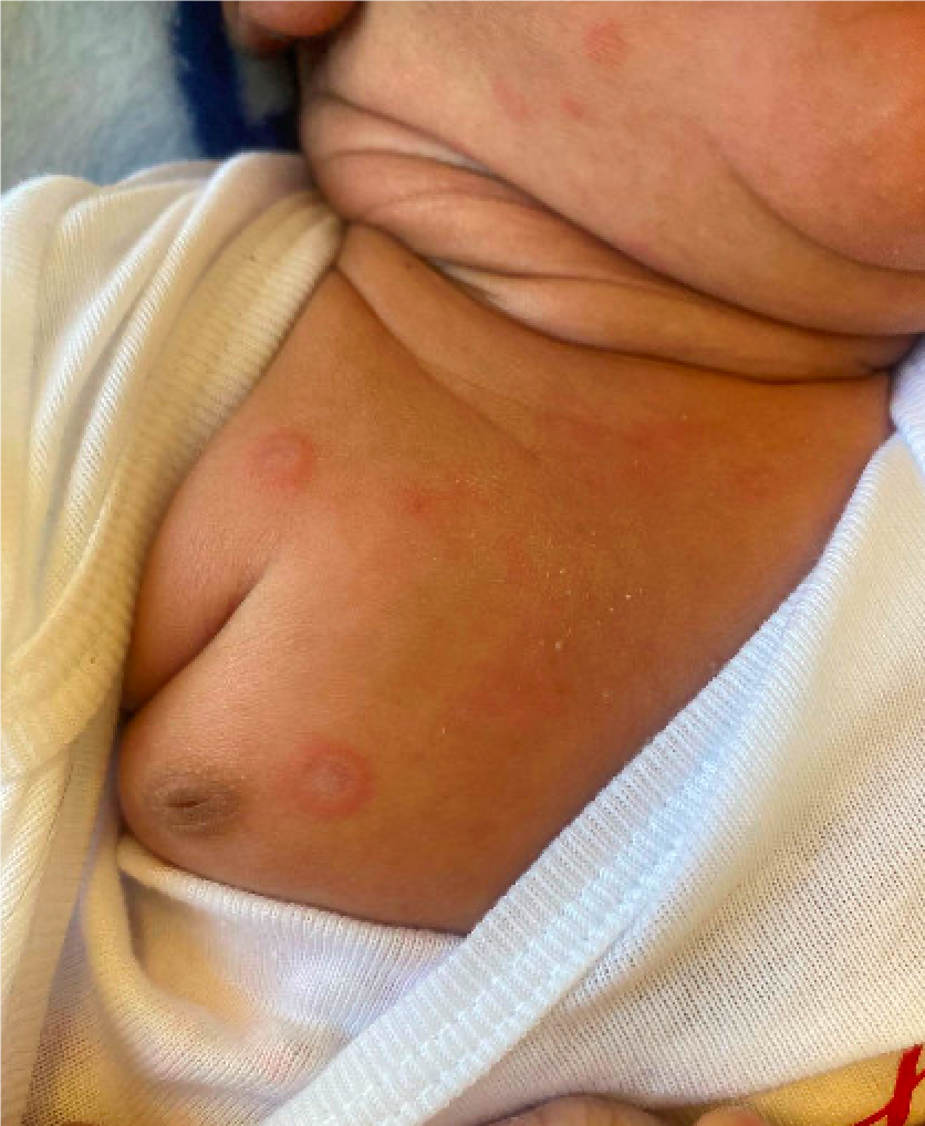
A 3-week-old infant presents to the dermatology clinic for evaluation of a rash. The rash developed around one week of life. Mother reported that the rash looked like “rings” and started on the scalp and face before spreading to the chest and abdomen.
The patient was born at 37 weeks. Labor was induced at that time due to maternal hypertension. The patient was diagnosed prenatally with abdominal heterotaxy, levocardia and small ventricular septal defects. Mother reported she was told the rash was due to allergic contact dermatitis to electrode adhesive from an electrocardiogram (ECG) that was performed during admission. The patient continued to develop new lesions after discharge prompting referral to dermatology by the patient’s pediatrician.
Which of the following is the most likely diagnosis?
- A) Dermatophyte infection (tinea capitis/tinea corporis/tinea faciei)
- B) Allergic contact dermatitis
- C) Neonatal lupus erythematosus
- D) Urticaria
- E) Erythema multiforme
Answer: C) Neonatal lupus erythematosus
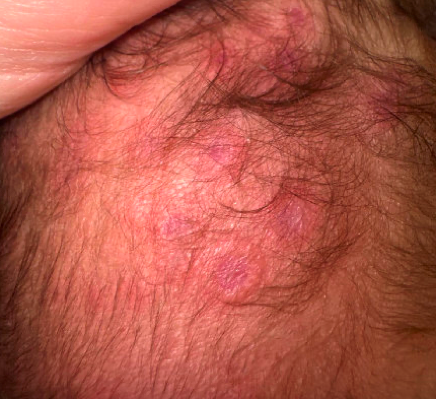
On examination, the patient has pink annular plaques with central clearing involving the scalp, face, chest and abdomen. The plaques are notable for an absence of scale. An annular non-scaly eruption in a newborn should raise suspicion for neonatal lupus erythematosus (NLE). NLE is important to recognize given the potential for systemic complications in the patient, as well as implications for the patient’s mother and for future pregnancies.
Skin manifestations of NLE are present at birth in two-thirds of patients; the remainder appear by the age of 5 months, although new skin lesions after 3 months of age are unusual. The most frequent site of involvement is the face but any body surface may be affected, and the rash may develop in sun-exposed areas. A variety of cutaneous manifestations of NLE can occur, including pink to red to hyperpigmented annular plaques with central clearing that are not typically scaly. Periorbital erythematous patches (“raccoon eyes”), discoid lesions (with atrophy and dyspigmentation), scaly atrophic macules and patches, and telangiectasia have also been described.
A dermatophyte infection (ringworm) is also on the differential diagnosis for annular lesions in newborns and infants. However, the annular plaques of tinea corporis usually have a peripheral rim of scale, not typically seen in NLE. A dermatophyte culture performed on a scraping from one of the lesions could also be considered.
Allergic contact dermatitis can occur in newborns but would be unlikely to present with annular plaques of varying shapes and sizes. In this case, the patient was initially diagnosed with allergic contact dermatitis to ECG electrode adhesives but had developed several lesions where leads would not have been placed and continued to develop new lesions long after the ECG had been performed.
Urticaria can be annular, but individual lesions typically resolve in 24 hours, whereas the lesions of NLE are fixed, lasting days to weeks before resolving.
While erythema multiforme can present with annular lesions, it would be unusual to occur in a newborn. In addition, the lesions of erythema multiform typically demonstrate concentric circles consisting of a well-circumscribed erythematous border, pale middle zone, and a dusky center (also called the target lesion), which were not seen in this case.
Which of the following diagnostic tests would be the next best step in management?
- ECG
- Brain magnetic resonance imaging
- Chest X-ray
- Skin biopsy
Answer: A) ECG
The major manifestations of NLE are cutaneous and cardiac findings. NLE is the most common cause of congenital heart block. Atrioventricular (AV) block occurs in 15% to 30% of cases of NLE. The proposed mechanism of how this occurs is the transplacental passage of maternal autoantibodies into the fetal circulation, where they bind to fetal cardiac cells, resulting in autoimmune injury and secondary fibrosis of the AV node and its surrounding tissue. An ECG and echocardiogram are recommended in patients with suspected NLE. Patients with first- and second-degree AV block can progress to third-degree AV block. Third-degree AV block is typically permanent and most of these patients will require pacemaker insertion at some point in life. There was no evidence of AV block in this patient’s case.
A skin biopsy is usually not necessary to diagnose neonatal lupus but could be considered if there is diagnostic uncertainty,
Other imaging may be indicated depending on the patient’s symptoms, exam findings and lab work results.
Which of the follow autoantibodies are most likely to be positive in patients with NLE?
- Anti-U1RNP antibody
- Anti-Sm antibody
- Anti-La (SSB) antibody
- Anti-Ro (SSA) antibody
Answer: D) Anti-Ro (SSA) antibody
Anti-Ro antibodies are detected in 95% of cases of NLE and anti-La in 60%-80% of cases. A small subset of affected infants have neither Ro nor La antibodies, but instead have anti-U1RNP antibodies. Anti-U1RNP antibodies do not typically cause AV block. Anti-Sm antibodies are usually not detected in NLE. Breastfeeding has not been shown to alter antibody levels or affect the development of cutaneous lesions.
While much less common than cutaneous and cardiac involvement, other systemic complications include liver, splenomegaly, lymphadenopathy and hematologic abnormalities (leukopenia, thrombocytopenia, anemia). Neurologic complications include central nervous system vasculitis and stroke and hydrocephalus. Pulmonary involvement is very rare.
Additional lab work that is recommended in patients with suspected NLE to evaluate for systemic involvement include a complete blood count, serum chemistry including liver functions tests and a coagulation screen (including lupus anticoagulant and antiphospholipid antibodies).
One of the most important aspects of management of patients with NLE is appropriate evaluation and management of the mother.
Approximately 30% of mothers of infants with NLE have a connective tissue disease, most commonly SLE or Sjögren syndrome. Most, however, are asymptomatic. Mother should be referred for appropriate evaluation for connective tissue disease to rheumatology and their obstetrician notified given the high risk of recurrence of NLE in future pregnancies.
Appropriate treatment of NLE includes avoidance of sun exposure and treatment of systemic complications, which may necessitate administration of systemic corticosteroid therapy. Low- to mid-potency topical steroids or topical calcineurin inhibitors can be used to treat the cutaneous lesions, but lesions clear spontaneously, and the use of topical anti-inflammatory agents has not been shown to affect risk of residua.
The patient’s lab work was notable for a positive anti-Ro antibody, anti-La antibody and anti-nuclear antibody (ANA), which together with the patient’s cutaneous findings confirmed the diagnosis of NLE. The remainder of the lab work was reassuring without evidence of other systemic complications of NLE. Mother’s obstetrician was contacted to arrange rheumatology evaluation and counseling regarding management of future pregnancies. At follow-up the patient’s skin lesions were resolving with some post-inflammatory dyspigmentation but no evidence of scarring.
References:
- Vanoni F, Lava SAG, Fossali EF, et al. Neonatal systemic lupus erythematosus syndrome: a comprehensive review. Clin Rev Allergy Immunol. 2017;53(3):469-476.
- Diociaiuti A, Paone C, Giraldi L, Paradisi M, El Hachem M. Congenital lupus erythematosus: case report and review of the literature. Pediatr Dermatol. 2005;22(3):240-242.
- Levy R, Briggs L, Silverman E, Pope E, Lara-Corrales I. Cutaneous sequelae in neonatal lupus: a retrospective cohort study. J Am Acad Dermatol. 2020;83(2):440-446. PMID: 31626881. doi:10.1016/j.jaad.2019.09.083
- Buyon JP. Neonatal lupus: epidemiology, pathogenesis, clinical manifestations, and diagnosis. UpToDate. UpToDate; 2025. https://www.uptodate.com/contents/neonatal-lupus-epidemiology-pathogenesis-clinical-manifestations-and-diagnosis
- Buyon JP. Neonatal lupus: management and outcomes. UpToDate. UpToDate; 2025. https://www.uptodate.com/contents/neonatal-lupus-management-and-outcomes